
Anatomy Of Brain Mri Anatomical Charts & Posters
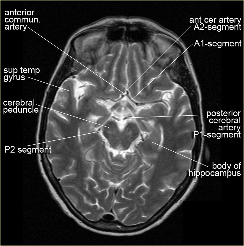
Also called ambient cistern is a cistern of the subarachnoid space between the posterior end of the corpus callosum and the superior surface of the cerebellum. It is sometimes defined as including the quadrigerminal cistern. On the left a coronal view of the segments of the middle cerebral artery. Horizontal M1-segment.
The Radiology Assistant Brain Anatomy
A brain (head) MRI scan is a painless test that produces very clear images of the structures inside of your head — mainly, your brain. Healthcare providers use brain MRIs to evaluate, diagnose and monitor several different medical conditions that affect your brain or other structures in your head.

Normal brain (MRI) Radiology Case Pediatric Radiology, Human Brain Anatomy
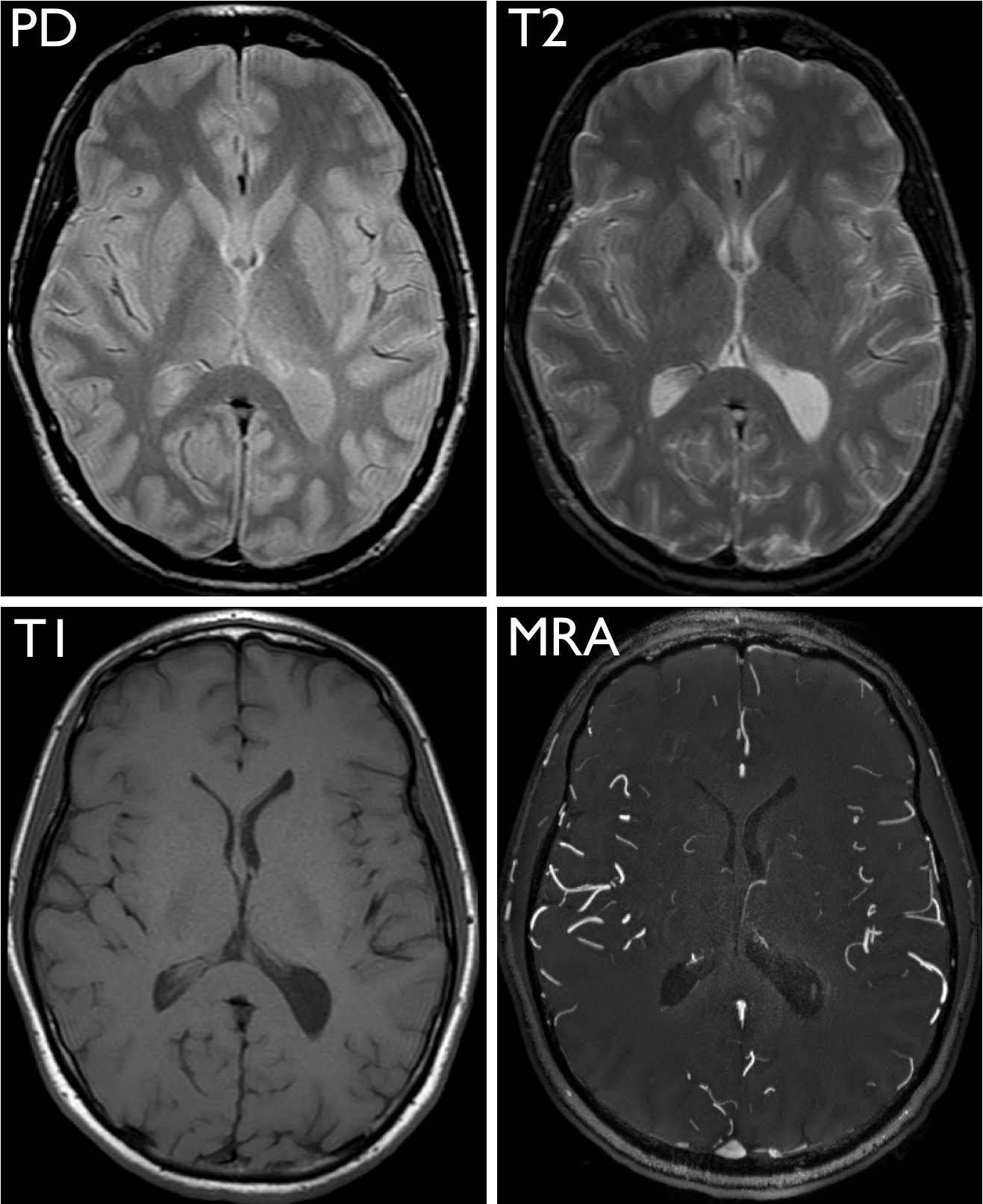
Brain magnetic resonance (MR) imaging studies provide multiple different imaging sequences in at least 2, and often 3, imaging planes. The different tissue signal characteristics and anatomic viewpoints are often complementary, and interpreting an MR imaging study of the brain can be a daunting task. The variety of pulse sequences and imaging.

Mri Anatomy Of Brain ANATOMY
MRI anatomy | Free MRI Axial Brain Anatomy AXIAL BRAIN SAGITTAL BRAIN CORONAL BRAIN CRANIAL NERVES ORBITS AND PNS TMJ CEREBRAL ARTERIES CEREBRAL VEINS NECK AXIAL NECK ARTERIES C SPINE AXIAL C SPINE SAGITTAL BRACHIAL PLEXUS CHEST AXIAL CHEST CORONAL HEART CHEST ARTERIES ABDOMEN AXIAL ABDOMEN CORONAL ABDOMEN ARTERIES BILIARY SYSTEM AXIAL
Brain Anatomy On Mri Anatomical Charts & Posters
A review of brain magnetic resonance imaging (MRI) is used as support. The anatomy of the brain is studied by means of axial, coronal and sagittal views. The MRI sequence used is a 3D gradient echo T1-weighted.
Approach to MRI brain
supporting tissues and structures The functional description of neuroanatomy divides the nervous system into: somatic nervous system autonomic nervous system This anatomy section promotes the use of the Terminologia Anatomica , the international standard of anatomical nomenclature 1. References Incoming Links Related articles: Anatomy: General
MRI Brain Anatomy
Sagittal 3D reconstruction Brain MRI with annotations of major structures. 13 articles feature images from this case 169 public playlists include this case

Brain MRI 3D normal anatomy eAnatomy
A special type of MRI is the functional MRI of the brain, also known as fMRI. It produces images of blood flow to certain areas of the brain. Functional MRI can be used to examine the brain's anatomy and show which parts of the brain are handling critical functions, language and movements. This information can help guide decisions when.

brain anatomy MRI coronal brain anatomy free MRI cross sectional anatomy Brain Anatomy
Brain anatomy Magnetic resonance imaging Sagittal Axial. Brain magnetic resonance (MR) imaging studies provide multiple different imaging sequences in at least 2, and often 3, imaging planes. The different tissue signal characteristics and anatomic view-points are often complementary, and interpreting an MR imaging study of the brain can be a.
Exploring the Brain How Are Brain Images Made with MRI? UCSF Radiology
To book a class, come to my website: https://www.alisanatomycourse.comThis video demonstrates the anatomy of the brain on MRI. It continues with a live inter.

S is for Syringomyelia This condition is the most common issue associated with Chiari. Do you
Brain Anatomy Content Reviewed: 2021-12-08 Preview Course View Contents Mastery Series is included with Membership Learn alongside world-renowned radiologists with interactive Mastery Courses. Watch expert-led lectures and case reviews from anywhere with short, bite-sized videos and practice on fully scrollable cases.

MRI anatomy brain axial image 14 Brain anatomy, Radiology, Mri brain
This video shows the appearance of the anatomical structures of the brain on a Magnetic Resonance Imaging.It aims to complement your understanding of neuroan.
Brain Anatomy On Mri Anatomical Charts & Posters
Anatomy of the brain (MRI) - cross-sectional atlas of human anatomy Antoine MICHEAU, MD , Denis HOA, MD Authors affiliations Publication date: Aug 25, 2008 | Last update: Oct 5, 2022 https://doi.org/10.37019/e-anatomy/163 ISSN 2534-5079

Delaware Neuroscience Brain Bee Detail, Page 2
The Whole Brain Atlas Keith A. Johnson, M.D. J. Alex Becker, Ph.D. Neuroimaging Primer - Harvard Medical School lecture notes: Introduction to Neuroimaging Normal Anatomy in 3-D with MRI/PET (Javascript) (Old) Atlas Navigator (Java) Normal Brain: Normal Anatomy in 3-D with MRI/PET (Javascript) Atlas of normal structure and blood flow

Brain Anatomy On Mri Anatomical Charts & Posters
Anatomical MRI has enabled systematic studies of brain development 16,17, ageing 18,19 (Fig. 1e), neurological disorders 20,21, trauma-related changes 22, and learning and plasticity 23,24,25,26.

Pin by SangHun, Jeong on Medical pictures Brain anatomy, Mri brain, Brain images
The MRI is a particularly powerful exam for studying structures such as diencephalon, mesencephalon (mid brain), pons, myelencephalon (medulla oblongata, bulb) and spinal cord. The vertical left menu provides reference images on coronal and sagittal views of the brain, with anatomical schemas based on a three dimensional (3D) model.